AJNS
CASE REPORT / CAS CLINIQUE
 
BROWN TUMOR OF LUMBAR SPINE IN PATIENT WITH CHRONIC RENAL FAILURE

LOCALISATION LOMBAIRE D'UNE TUMEUR BRUNE DANS L'INSUFFISANCE RENALE CHRONIQUE


  1. Department of neurosurgery, Ibn Rochd, University Hospital, Casablanca, Morocco
  2. Department of nephrology, Ibn Rochd, University Hospital, Casablanca, Morocco
  3. Laboratory pathological anatomy, Ibn Rochd, University Hospital, Casablanca, Morocco

E-Mail Contact - BERTAL Abderrazzak : abbertal@yahoo.fr


ABSRACT

Brown tumors are erosive bone lesions caused by increased osteoclastic activity. They usually occur in the severe forms of secondary hyperparathyroidism, as in patients with hemodialysis-dependent chronic renal disease. Involvement of the lumbar spine with this tumor causing neural compression is extremely rare. We report a 49-year-old man, who had been on haemodialysis for CRF for over 15 years, presented with leg weakness and back pain over the thoracolumbar junction. There were no motor or sensory disturbances. Spinal MRI revealed osteolytic lesions of the ribs and lumbar vertebrae L1. The clinical and radiological abnormalities resolved after parathyroidectomy and spine surgery.

Key-words: Brown tumor, hyperparathyroidism, lumbar spine, osteoclastomas, parathyroidectomy.

INTRODUCTION

Brown tumors or osteoclastomas are benign lesions of the bone, representing a reparative cellular process secondary of an hyperparathyroidism induced osteoclastic activity [1]. In patients with hemodialysis-dependent chronic renal disease, these lesions occur in the severe forms of secondary hyperparathyroidism. It occur commonly in the pelvis, ribs, clavicles and extremities, involvement in the spine is exceedingly rare [5]. The authors report a case of a 49 year-old man, submitted to a long period of hemodialysis, and who presented with a brown tumor of the lumbar spine, treated successfully.

CASE REPORT

A 49-year old man with a 15-year history of periodic hemodialysis-dependent chronic renal failure of unknown cause. He was referred to our department of neurosurgery for a back pain and a progressive bilateral lower-extremity weakness. On physical examination, there was mild tenderness over his lumbar spine. There were no motor or sensory disturbances, muscle tone was normal. Deep tendon reflexes and plantar responses were flexor, and sphincter tone was normal. Laboratory results demonstrated that serum levels were remarkable for creatinine of 12 mg/dl (normal range 0.6-1.2 mg/dl), for calcium of 11 mg/dl (normal range 8.6-10.5 mg/dl) and for PTH level of 1200 pg/ml. Plain radiographs and spinal MRI revealed osteolytic lesion of L1 lumbar vertebrae, causing the spinal cord compression (fig 1). The preoperative differential diagnosis included a metastatic tumor and a spinal tuberculosis.
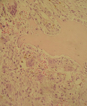
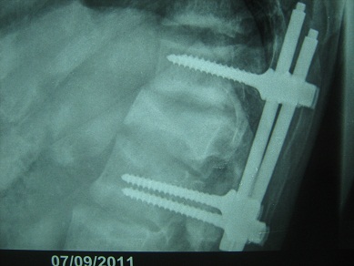
The Patient underwent a spinal surgery with total L1 laminectomy, transpedicular biopsy and lumbar fixation (fig 2). Postoperative Course was normal, the patient did well and was discharged five days later. Histological examination revealed hypercellular bone marrow with focal areas of fibrosis, multinucleate giant cells suggesting a brown tumor (fig 3). Two months later, the patient underwent a total parathyroidectomy, which normalized his serum PTH and calcium levels. Nine months postoperatively he was free from symptoms. The patient experienced an uneventful recovery and up to one year later, radiograph control showed the stability of instrumentation (fig 4), no local or distal recurrence was detected.

DISCUSSION

Brown tumors (BTs) or osteoclastomas also named osteitis fibrosa cystic are benign lesions of the bones. They correspond to a reparative cellular process secondary to an hyperparathyroidism-induced osteoclastic activity [1]. Bone defects are filled with fibroelastic tissue, which deforms the bone and simulates a neoplasm [9]. BTs arise as complications of a primary (3-7%) or a secondary hyperparathyroidism (1-2%), and the latter is a well-recognized complication in patients with end-stage renal failure, resulting in renal osteodystrophy including osteosclerosis, brown tumor and osteomalacia [8].
Brown tumors usually develop in the third to fourth decades of life, and females are more frequently affected [6]. It can occur as solitary or multiple lesions in any bone, the incidence of skeletal BT in patients with chronic renal failure (CRF) ranges from 1.5 to 13%, these lesions usually affect the pelvis, ribs, clavicles, mandible and extremities [8]. Involvement of the vertebral column is rare and mostly reported in thoracic spine. Since the first report case of a brown tumor involving the spine published in 1978 [2], very few cases were reported in the literature. The lumbar localization is considered exceedingly rare [6]. Gheith [2] reported in 2005, eight cases of spinal BTs, six cases were in thoracic and 2 cases located in the cervical spine. The present case is the second spinal BT reported in our department, the first one was located in the sacral region.

On neuroimaging, BTs realize lytic lesions that stimulate a little reactive bone formation, eventually misleading the diagnosis, especially when the lesions are multiples like in a secondary malignant tumor, amyloidosis, giant cell tumor [1,7,8] and spinal tuberculosis, which is still common in Morocco. Magnetic resonance imaging is very helpful in the accurate evaluation of the location and the extent of the tumor, exhibiting a low signal intensity on T1 and T2 weighted images, with foci of increased intensity corresponding to spots of hemorrhage [3,7]. However there is no specific image characteristics described for spinal BTs. Laboratory findings in patients with brown tumor include elevated serum levels of calcium and alkaline phosphatase and low serum levels of phosphate. The concentration of parathyroid hormone in serum is elevated.
Brown tumors are treated primarily by treating the underlying cause of the hyperparathyroidism and by using different management strategies for the bone lesions. It can be successfully treated by a parathyroidectomy and immobilization [1]. Spinal reconstruction in patients with CRF is controversial because of the severe osteoporosis and impaired bone healing [5]. However, when BTs involves the spinal cord, they can cause slowly progressive symptoms or acute compression secondary to bone fracture or by progressive features subsequent to the mass effect [2]. At this time, emergency surgical decompression of the spinal cord and instrumentation are required, mostly associated with parathyroidectomy, allowing stabilization of vertebral column and remineralization of the bone [4]. When patients have non progressive neurologic symptoms or multiple lesions without compression, surgical resection may not be necessary, biopsy of the lesion is essential for a definitive diagnosis, considering the rarity of brown tumors and the involvement of the spine. In this cases percutaneous CT-guided Tru-Cut needle biopsy of the vertebra is a safe and rapid method of an ultimate diagnosis. The histopathological features of brown tumor in hyperparathyroidism are characterized by a reduction in the number of trabeculae with local accumulation of osteoclastic multinucleated giant cells in fibrovascular stroma [1].The symptoms may disappear and remineralisation may take place with parathyroidectomy only [1,6].

CONCLUSION

In conclusion, BTs represent a reparative cellular process rather than a neoplastic process. Spine localisation, although rare, should be considered in the differential diagnosis of spinal lesions. Serum phosphate, calcium and PTH levels should be included in routine survey for multiple osteolytic lesions especially in patient with chronic renal failure. BTs can be resolves after parathyroidectomy, however surgical resection and stabilisation of spine may be needed to preserve neurologic function.
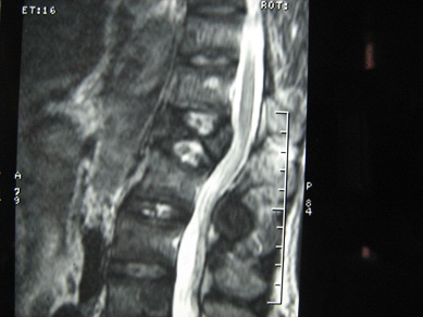

Figure 1
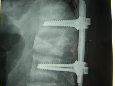
Figure 1


Figure 2

Figure 2


Figure 3

Figure 3


Figure 4

Figure 4


REFERENCES

  1. FINEMAN I, JOHNSON JP, DI-PATRE PL, SANDHU H. Chronic renal failure causing brown tumors and myelopathy. Case report and review of pathophysiology and treatment. J. Neurosurg 1999;90(2 Suppl):242-46.
  2. GHEITH O, AMMAR H, AKL A, HAMDY A, EL-SAEED M, EL-SALAMOUNY T, ADEL BAKER M, GHONEIM M. Spinal Compression by Brown Tumor in Two Patients With Chronic Kidney Allograft Failure on Maintenance Hemodialysis. Iranian Journal of Kidney Diseases 2010;4(3):256-9.
  3. HADDAD FH, MALKAWI OM, SHARBAJI AA, JBARA IF, RIHANI HR. Primary hyperparathyroidism. A rare cause of spinal cord compression. Saudi Med J 2007;28(5):783-86.
  4. JACKSON W, SELTHI A, CARP J, TALPOS G, VAIDYA R. Unusual spinal manifestation in secondary hyperparathyroidism: a case report. Spine (Phila Pa 1976) 2007;32(19):557-60.
  5. KAYA RA, CAVUSOGLU H, TANIK C, KAHYAOGLU O, DILBAZ S, TUNCER C, AYDIN Y. Spinal cord compression caused by a brown tumor at the cervicothoracic junction. Spine J 2007;7(6): 728-32.
  6. MAK KC, WONG YW, LUK KD. Spinal cord compression secondary to brown tumour in a patient on long-term haemodialysis: a case report. J Orthop Surg (Hong Kong). 2009;17(1):90-5.
  7. PADERNI S, BANDIERA S, BORIANI S. Vertebral localization of a brown tumor: description of a case and review of the literature. Chir Organi Mov 2003;88(1):83-91.
  8. RESIC H, MASNIC F, KUKAVICA N, SPASOVSKI G. Unusual clinical presentation of brown tumor in hemodialysis patients: two cases reports. Int Urol Nephrol 2011;43(2) :575- 80.
  9. TARRASS F, AYAD A, BENJELLOUN M, ANABI A, RAMDANI B, BENGHANEM MG, ZAID D. Cauda equina compression revealing brown tumor of the spine in a long-term hemodialysis patient. Joint Bone Spine 2006;73(6):748-50.


© 2002-2018 African Journal of Neurological Sciences.
All rights reserved. Terms of use.
Tous droits réservés. Termes d'Utilisation.
ISSN: 1992-2647