
|
|
|
CASE REPORT / CAS CLINIQUE
A RARE NEURONAL TUMOR OF THE CEREBELLUM WITH MYOID FEATURES
E-Mail Contact - CHBANI Laila :
chbanil@yahoo.fr
ABSTRACT We report an extremely rare tumor presenting with myoid features in the left cerebellar hemisphere in a 62-year-old man. This tumor consisted of medium to large round cells with focal lipomatous and myoid differentiation. Immunohistochemically, the tumor cells expressed synaptophysin, GFAP (glial fibrillary acidic protein) and focally desmin. From these findings, we concluded that this tumor was a liponeurocytoma with myoid features. To the best of our knowledge, this report constitutes the second described case of liponeurocytoma with myoid differentiation in the cerebellum. Keywords: liponeurocytoma, cerebellum, myoid differentiation, immunohistochemistry INTRODUCTION Cerebellar liponeurocytoma is morphologically composed of uniform round cells with neuronal differentiation and lipomatous features. It typically affects adults and has a favourable prognosis. Initially described as a lipomatous medulloblastoma, the World Health Organisation Working group proposes the term “cerebellar liponeurocytoma” primarily to distinguish this entity from medulloblastoma and to avoid over treatment of this benign tumor. CASE SUMMARY The patient was a 52-year-old man who presented with a 4-month history of headache of gradual onset, vomiting and decreased visual acuity. His past medical and family history were unremarkable. Neurological examination revealed cerebellar syndrome. Magnetic resonance imaging (MRI) showed a relatively well-defined, poorly enhancing, round lesion in the left cerebellar hemisphere hyperintense on T2-Weighted Images, causing moderate mass effect [Figure1]. At surgery, a 5,7cm x 4,7cm, well circumscribed mass, located in the cerebellar left hemisphere was removed. Ten percent formalin-fixed specimens were paraffin-embedded, cut at 3 µm, and stained with eosin-hematoxylin-saffron. Microscopically, the tumor was composed of small round cells to polygonal with eosinophilic or clear cytoplasm resembling oligodendrocytes [Figure 2]. In some areas, the tumor cells were mixed with lipomatous cells [Figure 3] and myocyte-like cells [Figure 4]. The tumor shows no mitotic figures. An immunohistochemical study was performed. After heat-induced epitope retrieval, dewaxed 3µm sections were incubated with the following antibodies: synaptophysin (Cell Marque, Rabbit polyclonal), GFAP (glial fibrillary acidic protein) (Cell Marque, Clone G-A-5, Mouse monoclonal), Mib1 (Cell Marque, Clone sp6, Rabbit monoclonal) and desmin (Cell Marque, clone D33, Mouse monoclonal). Revelation was performed after a streptavidin-biotin-peroxidase technique with diaminobenzidine. Tumour cells expressed synaptophysin [Figure 5], GFAP [Figure 6] and focally desmin [Figure 7]. The Mib 1 antibody stained less than 1% of tumour cell nuclei. Her postoperative period was uneventful with no evidence of tumour recurrence after 12 months. DISCUSSION Liponeurocytoma is a rare cebellar neoplasm of adults that is histologically characterized by neuronal differentiation with focal lipidization and low proliferative potential. Since the first report [2] by Bechtel in 1978 as a mixed mesenchymal and neuroectodermal tumor of the cerebellum, several terms “medullocytoma” [3], “lipomatous glioneurocytoma”[4] and “lipidized mature neuroectodermal tumor of the cerebellum” [5] have been proposed as to emphasize its resemblance to central neurocytoma. In 2000 [1], the WHO classification of tumours of the central nervous system proposed the term “cerebellar liponeurocytoma” to distinguish this tumor from medulloblastoma because of the outcome and to avoid aggressive treatment to this benign tumor. This tumor classically manifests at the fifth decades like our case (mean 50 years) [6] without significant gender predilection. This is in contrast with the age distribution of medulloblastoma. The clinical features are non-specific. This tumour is predominantly located in the cerebellar hemispheres like our observation followed by the vermis. Histologically, the cerebellar liponeurocytoma features a monotonous pattern of round cells with focal lipidization and neuronal differenciation reflected by immunoreactivity to neuronal markers [1]. The Mib1labelling index is even lower. In the literature data, only one case has been reported with additional immunoreactivity to desmin [5] in a 67 year-old woman. This myogenic feature hasn’t anysignificance. The most important differential diagnosis is medulloblastoma but the occurrence of the latter in children and the high Mib-1 labelling index can distinguish between these two tumors. The histogenesis of cerebellar liponeurocytoma has remained unclear. The immunoreactivity to neuronal antigens suggests that the fat containing cells result from lipomatous differentiation of tumour cells. The cell of origin is a precursor cell with commitment to neuronal differentiation but a capacity for divergent astrocytic and myogenic differentiation. Some studies suggests that this tumor is derived from the external granular layer of the cerebellum. [7] A complete surgical resection is recommended as the initial treatment but there is no consensus regarding the treatment of this rare tumor specifically adjuvant chemo-or radiotherapy. The prognosis of liponeurocytoma is favourable [8] CONCLUSION In conclusion, the cerebellar liponeurocytoma is a histopathologically and genetically distinct entity. The distinction between liponeurocytoma and medulloblastoma is very important since the medulloblastoma requires adjuvant radio/chemotherapy. 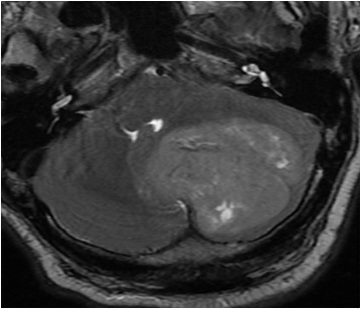 Figure 1: T2-Weighted MRI of cerebellar tumour. 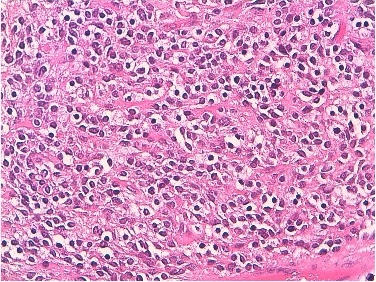 Figure 2: Hematoxylin-eosin-saffron stain x 250: tumour cells showing a round nuclei with a clear cytoplasm resembling neoplastic oligodendrocytes. 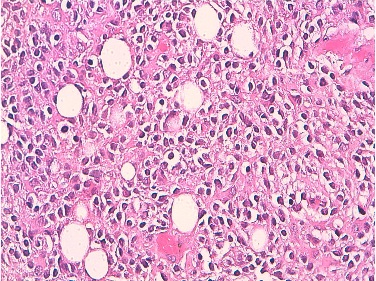 Figure 3: Hematoxylin-eosin-saffron stain x 250: Isomorphic small cells with focal lipomatous differentiation. 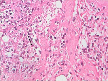 Figure 4: Hematoxylin-eosin-saffron stain x 250: myocyte-like cells (arrows) 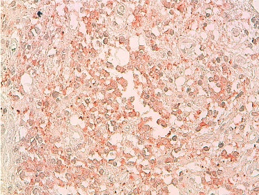 Figure 5: Immunoreactivity with anti-synaptophysin, DAB x 100. Small tumour cells express neuronal marker. 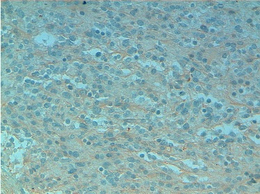 Figure 6: Immunoreactivity with GFAP 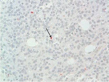 Figure 7: Immunoreactivity with anti-desmin, DAB x 100. Positivity of some tumor cells, indicating an myogenic differentiation. (arrows) REFERENCES
|
© 2002-2018 African Journal of Neurological Sciences.
All rights reserved. Terms of use.
Tous droits réservés. Termes d'Utilisation.
ISSN: 1992-2647