
|
|
|
NEUROANATOMY / NEUROANATOMIE
ANOMALOUS ORIGIN OF THE ULNAR NERVE FROM THE LATERAL CORD OF THE BRACHIAL PLEXUS: A CASE REPORT.
ORIGINE ANORMALE DU NERF ULNAIRE DU CORDON LATERAL DU PLEXUS BRACHIAL : A PROPOS D’UN CAS.
E-Mail Contact - MISIANI Musa :
tomamuti@gmail.com
ABSTRACT Background The ulnar nerve has been noted to bear variations as concerns its origin such as that of its communication with the median nerve via nerve branches and sharing of common sheath with the medial cutaneous nerve of the forearm. However, only a few studies have reported on the ulnar nerve receiving a communicating branch from the lateral cord. Case We report a case in which the ulnar nerve was noted to originate from branches of the lateral cord of the brachial plexus, during routine dissection. Discussion and conclusion This study adds data on such a rare scenario in a Kenyan setting. Keywords: Anomalous origin, Lateral cord, Ulnar nerve. RESUME Introduction Des variations de l’origine du nerf ulnaire telles que celle de sa communication avec le nerf médian via des branches nerveuses et le partage de la gaine commune avec le nerf cutané médial de l’avant-bras ont été décrites. Cependant, seules quelques études ont rapporté que le nerf ulnaire recevait une branche communicante du faisceau latéral. Observation Nous rapportons une observation dans laquelle le nerf ulnaire provenait des branches du cordon latéral du plexus brachial, lors d’une dissection de routine. Discussion et conclusion Cette étude ajoute des données sur un scénario rare dans un contexte kenyan. Mots-Clés : Cordon latéral, Nerf ulnaire, Origine anormale. INTRODUCTION Classically, the ulnar nerve originates as the continuation of the medial cord, with root values C8 and T1 following which it courses medial to the brachial artery until the insertion of coracobrachialis where it pierces the medial intermuscular septum from the anterior arm compartment to the posterior arm compartment (6). However, during routine dissection of the brachial plexus in our setting, a variation as concerns the origin of the ulnar nerve was noted. In the present case, the ulnar nerve was noted to originate from branches of the lateral cord of the brachial plexus. Following its origin the nerve coursed in its usual fashion, first medial to the brachial artery and then piercing the medial intermuscular septum. CASE REPORT During routine dissection of the brachial plexus, the ulnar nerve on the right side of a cadaver was noted to have a variant origin. It was carefully dissected till its origin from the brachial plexus, its origin and course noted and photographs taken. In our case, the cadaver had all the 3 cords of the brachial plexus. Interestingly, his right medial cord branched out to give the usual the medial pectoral nerve, medial cutaneous nerve of the forearm, medial cutaneous nerve of the arm, then later gave a branch that divided into two branches (termed upper and lower branch). Both branches appeared to be of equal thickness and length. The upper branch contributed in the formation of the median nerve whereas the lower branch (termed accessory ulnar nerve) joined the branch from the lateral cord termed (ulnar nerve proper). The other branches of the lateral cord were also noted: lateral pectoral nerve and musculocutaneous nerve. On the left side, the ulnar nerve was noted to originate classically as the continuation of the medial cord. DISCUSSION Previous studies on the origin of the ulnar nerve have noted variations however, few have been noted on its origin from the lateral cord of the brachial plexus (2,3). Among the common noted variations include its communication with the median nerve and its sharing of a common sheath with the median cutaneous nerve of the forearm (1). Fazan (2003) and Guru et al (2015) have also documented on the ulnar nerve receiving a communicating branch from the lateral cord (2,3). Usually such anomalous connections have been noted to result in anomalous innervation of intrinsic hand muscles in some cases, however in our case, the ulnar nerve was noted to course as usual and innervate its structures as described in the classical text. The basis surrounding the variant ulnar origin has been reported to be embryological, possibly due to the role of random factors influencing the mechanism of formation of limb muscles and peripheral nerves (5). Past research study has also described any communication between two nerves as a result of neurobiotaxis occurring during fetal development. Studies on the ulnar nerve variations in past research studies have reported that variant nerve sharing abnormal origin, course and distribution are more to accidental injuries and entrapment neuropathies especially during radical neck dissection, hence awareness of variations as such noted may be of surgical help. Further, unlike normal and anomalous positions of the arteries and the veins, which may be determined preoperatively by angiographic studies, it is not feasible to detect such in nerves especially owing to the fact that the current methods of detecting nerves such as Magnetic Resonance Imaging are currently a bit costly. As a result, knowledge of such variations may be highly important to anatomists, surgeons and clinicians alike (5).
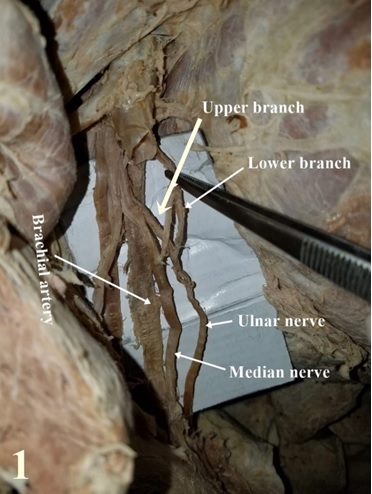 Figure 1: Figure showing the ulnar nerve receiving a branch from the lateral cord. REFERENCES
|
© 2002-2018 African Journal of Neurological Sciences.
All rights reserved. Terms of use.
Tous droits réservés. Termes d'Utilisation.
ISSN: 1992-2647