|
|
|
CASE REPORT / CAS CLINIQUE
COMPLETE INTRAVENTRICULAR MIGRATION OF A VENTRICULO-PERITONEAL SHUNT- A CASE REPORT AND BRIEF LITERATURE REVIEW
MIGRATION INTRAVENTICULAIRE D'UN SHUNT VENTRICULOPERIRONEAL
E-Mail Contact - KOMOLAFE Edward Oluwole :
eokomolafe@hotmail.com
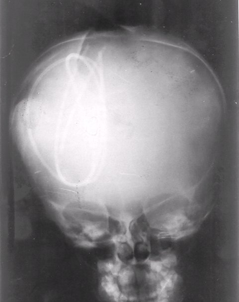
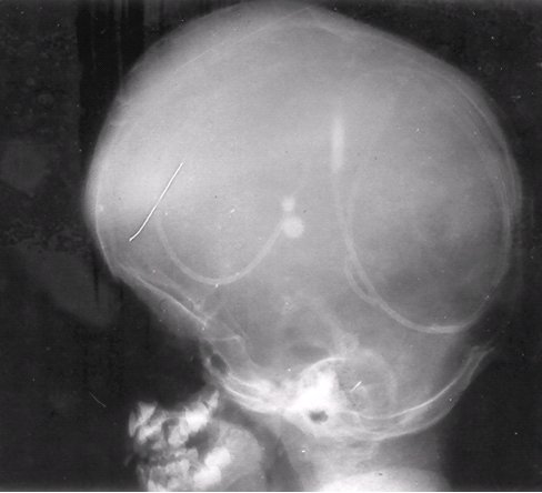
ABSTRACT Objects Methods Conclusion Keywords: Ventriculoperitoneal shunt, Hydrocephalus, Endoscopic third ventriculostomy, Shunt catheter. Mots cles: Complication, Hydrocéphalie, Intraventiculaire, Shunt ventriculo-péritonéal INTRODUCTION Cerebrospinal fluid (CSF) shunt operation was first realized in 1908 by Kausch [7] and since then ventriculoperitoneal (VP) shunts have remained the main method of treating hydrocephalus even with the recent increased interest in endoscopic third ventriculostomy. Although VP shunt procedures are easy to perform they are not without complications, adequate management of which may require further surgical procedures. These complications are many ranging from the very minor ones to major complications. Most often, despite appropriate surgical techniques, careful attention to operative details, strict compliance with asepsis and the use of prophylactic antibiotics to prevent or reduce the common complications that may follow VP shunt insertion, some complications still occur, some very unusual and rare [6]. One of these rare complications is complete proximal migration of the entire VP shunt system. Migration of the shunt or its parts have been reported but usually it is the distal or the peritoneal catheter that migrates after breakage or disconnection into many locations such as the scalp, heart, anus/rectum, urethral, knee, umbilicus, chest, pleural cavity, inguinal canal and scrotum [3]. Complete or total migration of the shunt as a whole is very rare and few cases are described in the literature. Distal migration rather than proximal shunt migration is more common and presumed to be due to intestinal peristalsis which may pull down the shunt. We aimed to present in this brief communication an unusual and a rare complication of Ventriculoperitoneal shunt placement which is total upward migration of the entire VP shunt system into the ventricular system of a child. The mechanism, management, and possible preventive methods are discussed. CASE REPORT AO is a 4year-old boy who first presented to our neurosurgical service at the age of five months with progressive hydrocephalus. VP shunt was advised but the parents refused until the child was 3years old due to poor milestone development and gross head enlargement. A right occipital VP shunt was inserted with satisfactory result. A year after, he presented with recurrent symptoms of active hydrocephalus and raised intracranial pressure associated with visual impairment. Examination revealed that the part of the VP shunt proximal to the shunt valve could not be palpated. This was confirmed by the shunt series, shuntogram, and computerized tomography (CT) brain scan which shows shunt disconnection proximal to the shunt valve with the ventricular catheter lying in the right lateral ventricle. There were no clinical features of sepsis and CSF culture did not yield any organism. A new shunt system was inserted with the aid of artery forceps leaving the detached ventricular catheter in situ. During the second shunt surgery, attention was placed on technical details so as to prevent recurrence of the complications, however the patient came back four weeks later with fever, vomiting, gross malnutrition and obtundation. This time no part of the VP shunt could be palpated along its entire length. A repeat shunt series shows the entire VP shunt system in the right lateral ventricle (Figure 1). CT scan was not done due to financial constraints. Sepsis screen did not yield any organisms but the patient was placed on, ceftriaxone. He later had limited craniectomy to retrieve the VP shunt system and the previously detached ventricular catheter. An external ventricular drainage was left in place to monitor and prevent excessive rise in the intracranial pressure until a new shunt could be placed. There was no improvement in his clinical and neurological status until three weeks later when his parent requested for discharge against medical advice. He presented once to the surgical out-patient clinic but had since been lost to follow-up. DISCUSSION Complete proximal migration of the entire VP shunt system is a well known complication though very rare in occurrence as few cases had been reported in the literature [10, 5]. This upward migration involves patient motion that creates a “windlass” effect with no resistance to the movement of the tubing and also requires a potential space such as the subgaleal or the ventricular spaces for the shunt to migrate to. Why this occurs in some patients is not known. A lot of factors had been attributed for the possible mechanism underlying this rare complication. These include the negative sucking intraventricular pressure, the positive pushing intraabdominal pressure and the tortuous subcutaneous track as well as neck movements. Other factors are related to the patient, the surgical technicalities and to the shunt itself. The patient related factors includes the age of the patient, severe and gross hydrocephalus with very thin cortical mantle, malnutrition, anaemia, sepsis, and repeated head movements and rotation (Bobble-head syndrome). Many of these predisposing factors were present in our patient who had a gross head enlargement with very thin cortical mantle. The patient presented also was malnourished and anaemic. The supine position in which the infants are nursed as well as the shorter distance between the ventricular and the peritoneal ends in these children facilitate proximal migration of the shunt. In all the cases reported in the literature all were infants and young children except Eljamel [5] that reported a case in an adult. This may not be unusual as majority of VP shunt are carried out in this age groups. In addition to the predisposing factors in this patient, we found out that the parents were constantly tampering with the shunt system at the region of the shunt valve, and thus likely to dislodge the shunt from the underlying tissues. This complication can be prevented by careful attention to surgical details particularly if performed by an experienced surgeon [1], careful patient selection, use of shunt with bulbous shunt valves and/or reservoirs, use of burr hole cover [4] to prevent the upward migration through the burr hole defect especially when large burr hole is made, and use of frontal burr hole site [3] with a small but preferably a cruciate rather than a linear dural incision to access the ventricle rather than other sites. The parents and/or the guardian of such children should also be educated on the care and handling to avoid undue tampering and dislodgement of the shunt system. The use of endoscopic third ventriculostomy (ETV) should be explored and encouraged for future treatment of hydrocephalus in selected and fit patients as an alternative to shunting procedures in selected patients [2].  Figure 1a  Figure 1b REFERENCES
|
© 2002-2018 African Journal of Neurological Sciences.
All rights reserved. Terms of use.
Tous droits réservés. Termes d'Utilisation.
ISSN: 1992-2647