|
||||||||||||||||||||||||||||||||||||||||||||||||||||||||||||||||
|
CLINICAL STUDIES / ETUDES CLINIQUES
INITIAL EXPERIENCE WITH NEUROENDOSCOPIC SURGERY IN WEST AFRICA.
EXPERIENCE INITIALE DE LA CHIRURGIE NEUROENDOSCOPIQUE EN AFRIQUE DE L'OUEST.
E-Mail Contact - ANDREWS Nii Adjetey Bonney :
neurogh@africaonline.com.gh
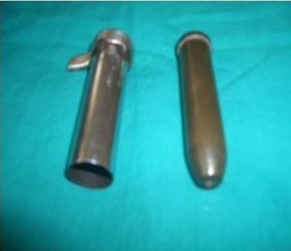
RESUME Introduction Patients and Methods Results RESUME Introduction Patients and Methodes Resultats Quatre-vingt huit pour cent des tumeurs intra-ventriculaires se présentaient sous forme d’une hydrocéphalie non communicante ; 57% présentaient une cécité. Deux de ces patients ont vu leur hydrocéphalie disparaître après la procédure neuroendoscopique. Deux autres ont nécessité une vérification externe suivi par une dérivation ventriculo-péritonéale. Trois patients ont eu une ventriculostomie. Deux patients – une sténose de l’acqueduc congénitale et en rapport avec un tumeur de la fosse postérieure – ont bénéficié d’une acqueducoplastie et d’une ventriculostomie. Un volumineux hématome putaminal d’origine hypertensive a été évacué. Conclusion Keywords : Afrique, endoscopie, hydrocéphalie, tumeurs cérébrales, ventriculostomie, Africa, neuroendoscopic surgery, endoscopic third ventriculostomy INTRODUCTION Neuroendoscopic surgery is commonly utilized for the management of intracranial cystic lesions (7, 16), hydrocephalus (9,18,27), tumor resections and biopsies (11,15,25) and for all types of microsurgical procedures that can involve endoscopic assistance, provided there is sufficient additional control of the operative field without retraction of neurovascular structures (7, 9). PATIENTS AND METHODS A retrospective audit of the first twenty patients who underwent neuroendoscopic procedures at our institution was performed. This was done by the evaluation of their clinical charts. The parameters examined were demographics, clinical, radiologic, operative and outcome data. Operative procedure. Statistical methods. 1)After a normal distribution test was applied, two sample t test was used to analyze which two groups are significantly different when compared to each other, p<0.05 was considered significant. 2)In order to compare two or more groups with outcome variables in more than two categories, a chi squared was used; where indicated, the Yates correction for continuity was applied, p<0.05 was considered significant. RESULTS Twenty patients (15M, 5F) with a mean age of 34 years (R 10months-74years) constituted the series (Table 1).They underwent a total of 23 neuroendoscopic procedures (Table 2). This constituted 29% of all intracranial procedures and 10% of all neurosurgical operations performed during the study period. Patient followup for the series averaged 17months (R 3-38months). In all instances preoperative diagnosis was by Head CT scan only. Stereotactic guidance was utilized in 13% (n=3) cases; all IVT. Eighty five percent (n=17) of the patients had a preop diagnosis of supratentorial tumor; 53% (n=9) were EVT; 47% (n=8) were IVT. A total of 48% of the neuroendoscopic procedures were performed at an extraventricular site. The histopathologic diagnosis of all tumors in the series was as follows : pituitary adenoma (5), ependymoma (5), astrocytoma (4), colloid cyst, craniopharyngioma, choroid plexus papilloma one each. Ependymoma was the commonest IVT (50%) and pituitary adenoma was the commonest EVT (55%). The sella was the location of 78% of the EVT (chi sq, p>0.05). Fifty percent of the tumors located in the sella presented with total blindness; 71% of the sella masses were pituitary adenoma (chi sq, p>0.05). The mean ages of pituitary adenoma and ependymoma patients were 47.4 (R40-50) years and 22.4 (R 5-41) years respectively; the difference was significant (t test, p<0.05). Seven (88%) of the patients with IVT presented with non-communicating hydrocephalus (NCHC) ; this was significant (chi sq, p<0.05). A total of 57% of these presented with total blindness. Two of the patients with IVT and NCHC underwent total neuroendoscopic tumor excision with complete resolution of hydrocephalus ; 2 required extraventricular drainage (EVD) followed by ventriculo-peritoneal shunting; 3 underwent subtotal resection followed by neuroendoscopic third ventriculostomy (NETV). There was one patient with congenital aqueductal stenosis and another with NCHC from a posterior fossa tumor; these patients underwent aqueductoplasty and NETV respectively. One patient underwent evacuation of a large hypertensive putaminal hematoma causing imminent herniation. The mean Karnofsky Performance Score (KPS) preop for IVT and EVT were 47 and 65 respectively; the difference is significant (t test, p<0.05). The postop KPS was calculated on the 14th postop day. No significant difference (t test, p>0.05) was found between i) preop and postop KPS for EVT, ii) post op KPS of IVT versus EVT. However, the KPS for IVT preop versus postop were significantly different (t test, p<0.05). The complications noted for the series within the first 28days after surgery were as follows : mortality rate of 4.4% and morbidity rate of 8.7%. One patient died on the 4th postop day following sudden onset of hemiplegia and mental status changes on the 2nd post op day following resection of a sella tumor. Another patient had a persistent CSF leak from a burr hole site after IVT resection. This resolved after duraplasty. DISCUSSION Our initial experience with neuroendoscopic surgery in West Africa is markedly different from that reported from East Africa (27). Neuro-oncologic applications dominate our series while the East African experience is marked by applications for the management of hydrocephalus. Neurooncology, in all its aspects, provides an ideal venue for the application of endoscopy (23). Eighty five percent of the patients in our series had supratentorial tumors; 47% of which were IVT with ependymoma (50%) being the commonest IVT. The advantages of improved visualization of intraventricular pathology, better management of tumor related hydrocephalus, less morbid biopsies and minimally invasive removal of IVT were invaluable adjuncts to traditional tumor management (23). We also did manufacture a prototype tubular retractor (“Sakumo Stereotactic Retractor”, Figure 4) which was adapted for stereotatic insertion into the ventricle or extraventricular compartment following frame based localization and the insertion of a stereotactic guiding needle. Consequently, we combined neuroendoscopy, microsurgery and stereotactic image guidance in our approach to 37% of IVT (6). With this technique, a rigid endoscope was used as a visualization tool, and microsurgical instruments were used for lesion removal via a 12mm conduit. The use of the prototype tubular retractor for access maintenance instead of conventional blade retraction provides several advantages. First, it leads to an even distribution of retraction forces. In contrast, conventional blade retraction localizes pressure at point of contact with brain parenchyma; this with IVT resection can cause damage to the caudate nucleus or internal capsule. Second, it allows for the use of microsurgical instruments that then enable tumors larger than 2cm to be resected. Conventional neuroendoscopy only allows for the placement of small and limited endoscopic instruments for lesion removal. Third, it allowed for the periodic reangulation of the endoscope, further enhancing the ability to resect larger IVT’s. Stereotactic targeting enabled us to gain safe access to other areas for which standard external landmarks are unreliable such as the posterior part of the third ventricle, atrium and occipital horns and the quadrigeminal cistern. The combination of stereotactic guidance, neuroendoscopy and microsurgery allowed us to successfully evacuate a large putaminal hemorrhage without prior angiography (Figure 2)! Fifty three percent of our tumors were EVT; 78% were in the sella and the commonest type was pituitary adenoma. Fully 50% of all the sella tumors presented with total blindness indicating late reportage and almost invariably large tumors with suprasella extension. This is reflected in the absence of significant difference in the KPS for EVT preop when compared to postop. Surgical treatment of large pituitary tumors with suprasellar extensions has been controversial. Both transcranial and transphenoidal approaches are sometimes far from satisfactory (2, 24). Recurrence rates have ranged from 20-42% when suprasellar extensions have exceeded 20mm (3, 17). Shanno et al (20) reported unchanged vision loss in 55% of their patients and Grade IV resections in 80% of their pituitary macroadenomas and all craniopharyngiomas. We elected to approach our sella lesions via a frontolateral craniotomy through a supraciliary incision; we then augmented this method by an endoscope-assisted microsurgical technique (12, 22, 13, 5). It is well worth emphasizing that the main purpose of the keyhole concept is not to diminish craniotomy size but to reconsider “standard’ craniotomy and subsequent intracranial procedure. The benefits of the suprabrow incision include shorter opening and closing times; better cosmetic results and a lower incidence of temporalis wasting. It also obviates the need for brain retraction and sylvian fissure dissection. Its disadvantages include difficulty harvesting pericranial graft and a potentially visible scar although the eyebrow usually obscures it. The use of the endoscope during tumor resection provided improved visual control of the retrosellar, endosellar, retrroclival and infratentorial structures in spite of the use of little or no traction. Transphenoidal surgery requires special training, special dedicated instruments and a good quality image intensifier. These resources are not readily available in our region. The case can therefore be made for pursuing keyhole approaches for the surgical treatment of the large pituitary tumors that often present in West Africa. Much earlier diagnosis of sella lesions is needed in order to reduce the number of patients presenting with total blindness and suprasellar extension. NETV and aqueductoplasty are currently the principal alternative to CSF shunt placement for the management of hydrocephalus (19). Shunts should and can be avoided whenever possible (27). A patient who undergoes a shunt procedure has a future life threatened by numerous complications and repeated operations. These dangers justify procedures that render the patient shunt independent especially since the cost of shunts readily available in West Africa is prohibitive. We had been reluctant to try NETV in patients with any indication of post infectious hydrocephalus (PIHC) hence the small number of NETV in our series, 15%. However the success achieved by Warf in Uganda in cases with PIHC (27) will encourage us to offer NETV to a greater number of our patients with different categories of hydrocephalus. CONCLUSION The initial experience with neuroendoscopic surgery in West Africa consists of the safe performance of IVT and EVT resections; NETV and aqueductoplasty for the management of hydrocephalus; and evacuation of intraaxial hematoma. The combination of neuroendoscopic surgery, stereotactic surgery and microneurosurgery has led to an expansion of minimally invasive techniques available for patient care in the region thus potentially improving patient outcomes and reducing complication rates.
We gratefully acknowledge the invaluable aid provided by A.A. Kelly, E. Andrews PE., Anton-Philip Battiade, J. Asamoah, C. Fiagah, C. Doku-Attuah, S. Bati, R. Ramesh, MD and E.N. Narh, MD TABLE 1
TABLE 2
 Figure 1 REFERENCES
|
© 2002-2018 African Journal of Neurological Sciences.
All rights reserved. Terms of use.
Tous droits réservés. Termes d'Utilisation.
ISSN: 1992-2647