
|
|
|
ORIGINAL PAPERS / ARTICLES ORIGINAUX
MULTIDISCIPLINARY NEURO-ONCOLOGY BOARD IN CÔTE D’IVOIRE: A FIVE-YEAR EXPERIENCE IN PEDIATRIC CENTRAL NERVOUS SYSTEM TUMOR CARE
STAFF DE NEURO-ONCOLOGIE EN CÔTE D’IVOIRE : CINQ ANS D’EXPÉRIENCE DANS LA PRISE EN CHARGE DES TUMEURS PÉDIATRIQUES DU SYSTÈME NERVEUX CENTRAL
ABSTRACT Background: Pediatric central nervous system (CNS) tumors are the second most common childhood cancer. In low-and middle-income countries, the management of these tumors remains challenging due to diagnostic delays and limited access to specialized care. Multidisciplinary tumor boards have emerged as an effective strategy to improve coordination of care and therapeutic decision-making. In Côte d’Ivoire, the introduction of radiotherapy enabled the establishment of the neuro-oncology tumor board in 2019. Objective: To describe the five-year experience of the Ivorian neuro-oncology board in the multidisciplinary management of pediatric CNS tumors. Methods: Retrospective longitudinal descriptive and analytical study of 53 patients aged 0-15 years with imaging-confirmed CNS tumors discussed at the neuro-oncology board between March 2019 and March 2024. Overall survival and recurrence-free survival were assessed using Kaplan-Meier method. Univariate analyses identified prognostic factors. Results: Mean age was 7.58±4.35 years with slight female predominance (sex ratio 0.89). Most patients (79.25%) had no health insurance. Diagnostic delay exceeded 3 months in 73.58% of cases. Gliomas were most frequent (49.05%), dominated by low-grade types (32.07%); followed by medulloblastomas (15.09%). Surgical treatment was performed in 90.6% of patients. One-year overall survival was 79.2% (95%CI: 69%-91%). Tumor type was significantly associated with survival (p=0.003), and complete resection prevented recurrence (p=0.017). Conclusion: This Ivorian multidisciplinary experience demonstrates encouraging results despite resource limitations. Major challenges include diagnostic delays, limited immunohistochemistry access, and lack of health insurance coverage. Strengthening this collaborative framework could reduce disparities with international standards. Keywords: Pediatric CNS tumors, Neuro-oncology, Multidisciplinary care, Low-resource setting
RÉSUMÉ Contexte : Les tumeurs du système nerveux central (SNC) représentent le deuxième cancer de l’enfant. La prise en charge est multidisciplinaire. En Côte d’Ivoire, l’introduction de la radiothérapie a facilité la création d’un staff multidisciplinaire de neuro-oncologie en 2019. Objectif : Décrire l’expérience du staff de neuro-oncologie ivoirien dans la prise en charge multidisciplinaire des tumeurs pédiatriques du SNC sur cinq ans. Méthode : Étude rétrospective longitudinale descriptive et analytique de 53 patients âgés de 0 à 15 ans, avec une tumeur du SNC confirmée à l’imagerie, discutés au staff de neuro-oncologie entre mars 2019 et mars 2024. La survie globale et sans récidive ont été évaluées par la méthode de Kaplan-Meier. Des analyses univariées ont identifié les facteurs pronostiques. Résultats : L’âge moyen était de 7,58±4,35 ans avec un sex-ratio à 0,89. La majorité des patients (79,25%) n’avaient pas d’assurance maladie. Le délai diagnostique dépassait 3 mois dans 73,58% des cas. Les gliomes étaient les plus fréquents (49,05%), particulièrement les bas grade (32,07%), suivis des médulloblastomes (15,09%). La chirurgie a été réalisée chez 90,6% des patients. La survie à un an était de 79,2% (IC95% : 69%-91%). Le type tumoral était significativement associé à la survie (p=0,003), et l’exérèse complète prévenait la récidive (p=0,017). Conclusion : Cette première expérience multidisciplinaire ivoirienne démontre des résultats encourageants. Les défis majeurs incluent le retard diagnostique, l’accès limité à l’immunohistochimie et l’absence d’assurance maladie. Le renforcement de ce cadre collaboratif est nécessaire. Mots-clés : Tumeurs pédiatriques du SNC ; Neuro-oncologie ; Prise en charge multidisciplinaire ; Ressources limitées Pediatric central nervous system (CNS) tumors represent the second most common childhood malignancy after leukemia and are the leading cause of cancer-related mortality in children (8,13). These tumors comprise a heterogeneous group of neoplasms with varied clinical, radiological and histopathological presentations, requiring multimodal treatments including surgery, chemotherapy, and radiotherapy according to tumor type (14,16). In high-income countries, advances in molecular diagnostics, neuroimaging, surgical techniques, and multidisciplinary collaboration have led to improvements in survival outcomes over recent decades (5,13). In contrast, outcomes in low- and middle-income countries (LMICs) remain poorer due to diagnostic delays, limited access to advanced imaging and histopathological examinations, insufficient specialized services and structured multidisciplinary care (19). Multidisciplinary tumor boards have emerged as a cornerstone of modern oncological care, providing a structured platform for collective discussion of complex cases by specialists from different disciplines (2,7). These platforms have demonstrated multiple benefits: reduction of delays between diagnosis and treatment initiation, improvement in the quality of therapeutic decisions for complex cases, standardization of care protocols, opportunities for continuing education for care teams, and systematic identification of patients eligible for clinical trials (9,15). In low and middle-income countries, innovative models such as telemedicine-based multidisciplinary meetings have demonstrated their feasibility and potential to improve pediatric neuro-oncology care (17,18). In Côte d’Ivoire, the introduction of radiotherapy facilitated the establishment of a multidisciplinary neuro-oncology tumor board in 2019, with the objective of harmonizing therapeutic decisions and improving patient care. Few studies have documented the experience of multidisciplinary management of pediatric CNS tumors in sub-Saharan African countries. To date, no publication has reported the results of such an initiative in Côte d’Ivoire. The present study aims to describe the five-year experience of the Ivorian neuro-oncology tumor board in the multidisciplinary management of pediatric CNS tumors. The specific objectives were to describe the organization and functioning of the neuro-oncology board; analyze the sociodemographic, clinical, radiological and histopathological characteristics of pediatric CNS tumors discussed; and evaluate one-year overall survival, recurrence-free survival, and associated prognostic factors. METHODS We conducted a retrospective longitudinal descriptive and analytical study of pediatric CNS tumor cases discussed over a five-year period, from March 1, 2019, to March 1, 2024. The study was conducted within the neuro-oncology tumor board of Côte d’Ivoire, a multidisciplinary consultation platform bringing together specialists involved in the management of central nervous system (CNS) tumors, including neurosurgeons, oncologists, neuro-oncologists, radiotherapists, radiologists, neuropathologist, neurologists, and pediatric oncologists. Since 2019, neuro-oncology tumor board meetings have been held every two weeks. Initially organized in person, meetings have been conducted via videoconference since the COVID-19 pandemic, allowing broader participation from specialists across different hospital centers nationwide. Participants originate from the Teaching Hospitals (CHU) of Yopougon, Cocody, Treichville, Bouaké, the Military Hospital of Abidjan, Saint Joseph Moscati Catholic Hospital in Yamoussoukro, and the National Center for Medical Oncology and Radiotherapy Alassane Ouattara (CNRAO). On average, twenty practitioners participate in each meeting. Decisions are made collegially by consensus after standardized presentation of each case including available clinical, radiological, and histopathological data. Patients were typically presented to the tumor board in the following situations: newly diagnosed CNS tumor requiring therapeutic planning, complex cases requiring multidisciplinary decision-making, postoperative cases requiring adjuvant treatment planning, suspected tumor recurrence or progression. When the minimum quorum of three cases is not reached, clinical discussions continue within a dedicated secure messaging group. This tumor board is not specifically pediatric, but regularly manages pediatric cases with occasional support from an external pediatric neuro-oncologist when necessary. The study population consisted of all pediatric patients aged 0 to 15 years whose CNS tumor cases were discussed during neuro-oncology tumor board meetings within the study period. Inclusion and Exclusion Criteria We included all patients aged 0 to 15 years with an intracranial or spinal tumor confirmed by imaging, whose cases were discussed at the neuro-oncology tumor board, and for whom a medical record was available. Patients with incomplete medical records preventing data exploitation and patients lost to follow-up were excluded from the study. Data were collected retrospectively from two main sources: neuro-oncology tumor board meeting records and patients’ hospital medical files. Tumor board meetings followed a standardized case presentation format, including clinical history, imaging findings, histopathological results when available, and proposed management strategies. All data were anonymized and entered into a standardized data collection form designed for the study. Variables Collected variables were: -Sociodemographic data: sex, age, health insurance coverage, personal and family medical history. -Clinical and imaging data: presenting symptoms, duration of symptom evolution, imaging modality used, tumor location -Histopathological data: tumor type and grade, and immunohistochemistry results -Therapeutic data: treatment modalities (surgery, radiotherapy, chemotherapy), type of surgery, extent of resection -Outcome data: tumor recurrence or progression and mortality within one year of diagnosis Data Analysis Data were analyzed using Microsoft Excel and R software (version 4.5.0). Qualitative variables were summarized as frequencies and percentages, while quantitative variables were summarized using means, medians, and standard deviations. Overall survival and recurrence-free survival at one year were estimated using the Kaplan–Meier method. The primary analytical objective was to explore the association between tumor type and one-year overall survival. Secondary exploratory analyses assessed associations between one-year survival and tumor location or age at diagnosis, as well as the association between extent of surgical resection and tumor recurrence or progression at one year. Univariate analyses were performed using Pearson’s chi-square test or Fisher’s exact test, as appropriate. Statistical significance was set at p < 0.05. Ethical Considerations We obtained authorization from the hospital administration. Data confidentiality was strictly maintained by assigning an anonymization number to each data collection form. There were no conflicts of interest. RESULTS During the study period, 53 pediatric CNS tumors were included. Female patients represented 53% of cases, with a sex ratio of 0.89. The mean age at diagnosis was 7.58±4.35 years. The majority of patients (69.81%) were aged 5-15 years. The majority of patients (79.25%, n=42) had no health insurance coverage at diagnosis. Only 11 patients (20.75%) had health insurance. Most patients had no specific medical and surgical histories, except for one patient followed for Wilms tumor who had undergone nephrectomy with chemotherapy. Regarding family history, only one case of familial cancer was identified: a first cousin who died of glioblastoma at age 25. Intracranial hypertension was the most common clinical presentation, observed in 37 patients (69.81%), followed by cerebellar syndrome in 9 patients (16.98%) and motor deficit in 7 patients (13.21%). Clinical symptoms had evolved for more than 3 months in 39 patients (73.58%), reflecting significant diagnostic delay. Thirteen patients (24.53%) presented with symptoms evolving for 2 weeks to 3 months. One patient (1.89%) had symptoms for less than 2 weeks. Magnetic resonance imaging (MRI) was the primary diagnostic imaging modality, performed in 43 patients (81.13%). Computed tomography (CT) was used in 10 patients (18.87%). No intramedullary tumors were identified. All cases involved intracranial tumors. The distribution between supratentorial (49%) and infratentorial (51%) locations was nearly balanced. Gliomas represented the most frequent tumor type with 26 cases (49.05%), dominated by low-grade forms (17 cases, 32.07% of all tumors) followed by high-grade gliomas (9 cases, 16.98%). Medulloblastomas constituted the second most frequent tumor type with 8 cases (15.09%). Supratentorial tumors were predominantly gliomas (12 cases), mainly low-grade gliomas (9 cases). Infratentorial tumors consisted primarily of medulloblastomas (8 cases) and low-grade gliomas (8 cases). The 17 low-grade gliomas included pilocytic astrocytoma (8 cases, 47%), diffuse low-grade glioma (6 cases, 35%), dysembryoplastic neuroepithelial tumor (2 cases, 12%), and ganglioglioma (1 case, 6%). Immunohistochemistry was performed in only 7 patients (13%); 46 patients (87%) did not undergo immunohistochemical analysis. Forty-eight patients underwent surgery (90.6%). Surgery alone was performed in 27 cases (56.25% of operated patients), and surgery followed by adjuvant treatment in 21 cases (43.75% of operated patients). The surgical procedure was gross total resection in 25 cases, partial resection in 14 cases, biopsy in 7 cases, and Ommaya reservoir placement with interferon injection in 2 cases. Adjuvant treatment consisted of postoperative radiotherapy (12 cases, 22.6%), concurrent chemoradiotherapy (8 cases, 15.1%), and postoperative chemotherapy alone (1 case, 1.9%). Radiotherapy without surgery was performed in 2 cases. Three patients received no specific treatment. This included two patients with high-grade glioma in whom radiotherapy was indicated but who died before treatment initiation, and one patient with cerebral lymphoma diagnosed on imaging who improved clinically after corticosteroid trial therapy without chemotherapy. At one year, 4 cases of recurrence were observed (8% of patients). These included two low-grade gliomas (oligoastrocytoma grade 2, oligodendroglioma), one supratentorial ependymoma in which postoperative radiotherapy was initiated after six weeks, and one hemangioblastoma treated with surgery alone. Overall survival at 12 months was estimated at 79.2% (95% CI: 69%-91%) with 11 deaths observed among the 53 included patients, corresponding to a mortality rate of 20.8% (figure 1) There were 7 high-grade glial tumors, 1 medulloblastoma, 1 rhabdomyosarcoma, 1 low-grade glioma (oligodendroglioma) that recurred and resulted in death one month after recurrence, and 1 ependymoma. The mean age of deceased patients was 8.54 years. Analyses were performed to explore associations between different variables and one-year survival, as well as between surgical procedure and tumor recurrence. Association between tumor type and one-year survival (table 4): Among the three most frequently encountered tumor types (low-grade gliomas, high-grade gliomas, and medulloblastomas), a statistically significant association was observed between tumor type and one-year survival (p=0.003; Fisher’s exact test). Low-grade gliomas were associated with higher survival (94%) compared to high-grade gliomas (67%). Association between tumor location and one-year survival (table 4): No statistically significant association was found between tumor location (supratentorial vs infratentorial) and one-year survival (p=0.36; Chi-square test). Association between age and one-year survival (table 4): No statistically significant association was found between age at diagnosis (<5 years vs ≥5 years) and one-year survival (p=0.736; Fisher’s exact test). Association between surgical procedure and recurrence/progression (table 5): Fisher’s exact test revealed a statistically significant association between the type of surgical procedure and occurrence of recurrence or tumor progression (p=0.017). No recurrence was observed in patients who underwent complete tumor resection. This study reports the first experience of a structured multidisciplinary neuro-oncology tumor board for pediatric CNS tumors in Côte d’Ivoire. Over a five-year period, this initiative allowed multidisciplinary discussion and management of 53 pediatric CNS tumor cases. This series provides valuable epidemiological and clinical data in an African context where such information remains scarce. Achievement of the study objectives The primary objectives of this study were successfully achieved. We described the organization and functioning of the Ivorian neuro-oncology tumor board, characterized by regular meetings, broad multidisciplinary participation, and nationwide collaboration through videoconferencing. We also analyzed the sociodemographic, clinical, radiological, and histopathological characteristics of pediatric CNS tumors discussed within this framework, and evaluated one-year overall survival and recurrence-free survival, as well as associated prognostic factors. These findings provide valuable insight into pediatric neuro-oncology care in Côte d’Ivoire, where published data remain scarce. Organization of the multidisciplinary tumor board The establishment of the neuro-oncology tumor board in 2019 created a structured platform for multidisciplinary consultation, facilitating collegial discussions among specialists involved in pediatric CNS tumor management. The adoption of videoconferencing since the COVID-19 pandemic has maintained regular collaborative dynamics with an average participation of twenty practitioners per meeting from different hospital centers nationwide. This approach aligns with successful tele-mentoring experiences in pediatric neuro-oncology reported by Qaddoumi et al. in Jordan (17,18), demonstrating the positive impact of remote collaboration in improving quality of care in resource-limited contexts. Similarly, Foo et al. reported improved coordination and reduced management delays following the implementation of a neuro-oncology tumor board in Malaysia (4). Our findings confirm that such collaborative models are reproducible and relevant in sub-Saharan Africa. Epidemiological and clinical Characteristics With 53 patients over five years, our series represents approximately 10.6 cases annually. This figure, though modest, is consistent with data from other resource-limited countries and likely reflects underestimation of true incidence due to limited access to care and underdiagnosis (11,12). For comparison, the Malaysian series reported 12.25 cases/year (4), and the previous Ivorian series by Broalet et al. identified 5.2 cases/year between 1995-2006 (1) . The relative increase in detected cases may reflect progressive improvement in access to diagnostic imaging and healthcare professional awareness. Our study revealed female predominance (53%, sex ratio 0.89), consistent with the previous Ivorian series by Broalet et al.(1) which also reported marked female predominance. This Ivorian particularity contrasts with most international and some African series showing slight male predominance (11,12,19). The persistence of female predominance in two successive Ivorian series suggests either a local epidemiological characteristic or selection bias related to sociocultural factors influencing healthcare access differently by sex. Mean age at diagnosis (7.58 years) with predominance of the 5-15 year age group (69.81%) was comparable to other African series where mean age typically ranges between 6.5 and 9 years (6,11,12), reflecting the classic peak incidence of pediatric CNS tumors between 5 and 10 years. Diagnostic delay was a major challenge with 73.58% of patients presenting after three months of symptom evolution. Similar delays have been widely reported in sub-Saharan Africa and contribute to advanced disease at presentation and poorer outcomes (3,6). The absence of health insurance in 79.25% of patients constitutes a major barrier to equitable healthcare access. The cost of diagnostic examinations (multimodal MRI, histopathology), surgery, and adjuvant treatments represents catastrophic financial burden for families, frequently leading to treatment delays. This problematic is widely documented in sub-Saharan Africa and constitutes one of the main explanations for survival disparities compared to high-income countries (6,11,19). In Mittal et al.’s meta-analysis (11), the surgical rate in sub-Saharan Africa was only 77% versus 97.4% in North Africa, a difference largely attributed to financial barriers. Limited access to immunohistochemistry and molecular analysis: No molecular analysis is available in Cote d’Ivoire. Immunohistochemistry was performed in only 13% of our patients. This is explained by the unavailability of immunohistochemical explorations in Côte d’Ivoire before 2021, as well as their high cost even after introduction. This situation is common to most sub-Saharan African countries. Implementation of the 2021 WHO classification (8), which integrates essential molecular parameters, remains a major challenge in sub-Saharan Africa where diagnosis still relies largely on conventional histopathology. Tumor Characteristics Our series was distinguished by a high proportion of low-grade gliomas (32.07%), particularly pilocytic astrocytomas (15% of all tumors), consistent with data from Stagno et al. (19) and suggesting that these slow-growing tumors are more frequently detected despite diagnostic delays, while some very aggressive tumors might never reach specialized centers. The distribution between infratentorial (51%) and supratentorial (49%) locations was nearly balanced, consistent with classic pediatric data where approximately 60% of tumors are infratentorial (13). Treatment and Outcomes Surgery constituted the cornerstone of management in our series, performed in 48 of 53 patients (90.6%). This surgical rate is superior to the sub-Saharan African average of 77% reported by Mittal et al. (11), but inferior to North Africa 97.4%, explained by a combination of factors: availability of local pediatric neurosurgical expertise, but persistence of financial barriers and medical contraindications in some patients with very advanced presentations. Adjuvant treatment was dominated by postoperative radiotherapy (22.6%), followed by concurrent chemoradiotherapy (15.1%). Access to radiotherapy represents a major advance for Côte d’Ivoire. Previously, patients requiring radiotherapy had to be evacuated abroad, which was financially prohibitive for most families. Overall survival at one year was 79.2% (95% CI: 69%-91%), an encouraging result situated in the upper range of sub-Saharan African data. Broalet et al.’s Ivorian series reported 22.8% mortality (1), and Mittal et al.’s meta-analysis reported average mortality of 30.3% in Africa (36.7% in sub-Saharan Africa vs 18.8% in North Africa) (11). These results suggest that structured multidisciplinary care may contribute to improved outcomes, even in resource-limited settings. Prognostic Factors We identified significant associations between tumor type and survival (p=0.003), with low-grade gliomas showing excellent prognosis (94% survival) compared to high-grade gliomas (67%) and medulloblastomas. This result is clinically meaningful in pediatric neuro-oncology, where histological grade is a major determinant of short-term and long term outcomes We also demonstrated a statistically significant association between extent of surgical resection and recurrence (p=0.017), with no recurrence observed after complete resection. This result emphasizes the crucial importance of surgical radicality when anatomically and functionally feasible, consistent with international data establishing complete resection as a major prognostic factor in most pediatric CNS tumors (10,16). Contrary to some literature, we found no statistically significant association between tumor location (supratentorial vs infratentorial) and one-year survival (p=0.36), nor between age at diagnosis and survival (p=0.736). These negative results might be explained by histological diversity of tumors in each location and relatively limited sample size reducing statistical power. Study Limitations and perspectives Our study has several limitations: retrospective nature with incomplete medical records, limited follow-up duration (one year prevents assessment of long-term survival particularly important for low-grade tumors), and insufficient immunohistochemistry with absence of molecular analysis limiting diagnostic precision and tumor classification. The absence of molecular classification, as recommended by the 2021 WHO classification, remains a major challenge in Côte d’Ivoire and most sub-Saharan African countries. Despite these limitations, this study demonstrates the feasibility and relevance of structured multidisciplinary tumor boards in pediatric neuro-oncology care in resource-limited settings. Maintaining and strengthening this collaborative framework, combined with recommendations for improving access to care, developing a national registry, strengthening immunohistochemistry and molecular biology capacities, and establishing international partnerships for tele-mentoring, could contribute to progressively reducing disparities with international standards while advancing knowledge in African pediatric neuro-oncology. CONCLUSION This study reports the first experience of structured multidisciplinary management of pediatric CNS tumors in Côte d’Ivoire through a neuro-oncology tumor board established in 2019. Over five years, 53 patients were managed with encouraging outcomes: one-year overall survival reached 79.2% (95% CI: 69%-91%) and surgical treatment was achieved in 90.6% of cases. Our results demonstrate that multidisciplinary tumor board implementation is feasible and beneficial in resource-limited settings, with videoconferencing enabling effective collaboration across multiple centers nationwide. Despite these encouraging results, major challenges persist. Significant diagnostic delay, limited access to immunohistochemistry, and lack of health insurance coverage remain substantial barriers to optimal care. These findings underscore the critical need for strengthening healthcare infrastructure and improving access to specialized diagnostic and therapeutic resources in our context. Our study identified significant prognostic factors that should guide clinical practice: tumor type was significantly associated with survival (p=0.003), and complete surgical resection was associated with absence of recurrence (p=0.017), emphasizing the importance of maximal safe resection when feasible. These findings align with international literature and validate their applicability in resource-limited settings. The multidisciplinary tumor board model presented here is reproducible for other sub-Saharan African centers. Key success factors include regular meetings with broad specialist participation, videoconferencing to overcome geographical barriers, and collegial decision-making processes. This work contributes valuable data to the limited body of literature on pediatric neuro-oncology in sub-Saharan Africa and demonstrates that improved outcomes are achievable through multidisciplinary collaboration, even in resource-constrained environments. AUTHOR CERTIFICATION I, the undersigned, Doctor Grace Ella Armande DJONDE, corresponding author of this manuscript, hereby certify that: -I assume full responsibility for the content of this manuscript. -All authors have made substantial contributions to this work and agree to its submission for publication in the African Journal of Neurological Sciences (AJNS). -All persons listed as authors have read and approved the final version of this manuscript and the mention of their names as authors. -This manuscript represents original work that has not been published elsewhere, in whole or in part, and is not currently under consideration by another journal. -All participants in this research have been treated in accordance with ethical standards, and all necessary authorizations have been obtained. 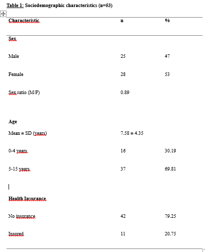 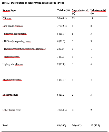 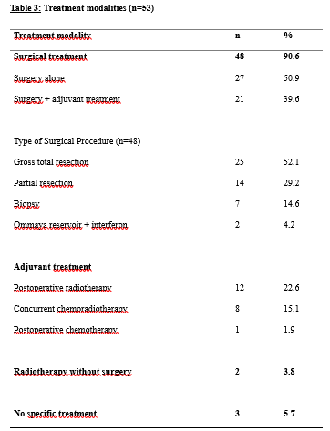 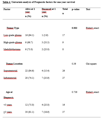 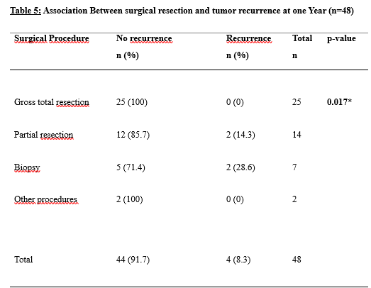 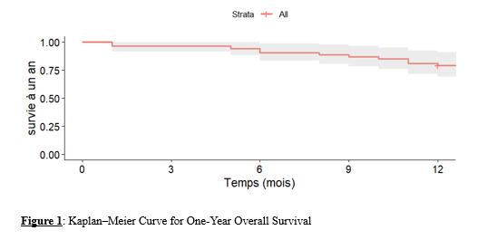 REFERENCES
|
© 2002-2018 African Journal of Neurological Sciences.
All rights reserved. Terms of use.
Tous droits réservés. Termes d'Utilisation.
ISSN: 1992-2647