CASE REPORT / CAS CLINIQUE
MULTIPLE INTRACRANIAL GERMINOMAS: A CASE REPORT
GERMINOME INTRACRANIEN MULTIPLE: A PROPOS D'UN CAS
- Department of neurosurgery, Ibn Rochd, University Hospital, Casablanca, Morocco
E-Mail Contact - CHELLAOUI Abdelmajid :
ABSTRACT
Germinomas are malignant tumors arising from primitive germ cells. They may be solitary or multiple and can be situated along the midline structures or in other intracranial regions. We report on one case of multiple intracranial germinoma. A 38 year-old male patient presented with signs of increased intracranial pressure and bilateral dimunition of visual acuity. A brain MRI showed multiple lesions at the level of the pineal region, the suprasellar region and in the right pontocerebellar angle. Tumor markers were positive. He underwent radiation therapy and chemotherapy with good recovery. To the best of our knowledge no similar case was described in the African literature before.
Key-words: Germinoma – Brain – Suprasellar – Pineal region
RESUME
Les germinomes sont des tumeurs malignes dérivant des cellules primitives germinales occupant les structures de la ligne médiane au niveau cérébral telle la région pinéale et/ou sellaire. Nous rapportons un cas de localisation multiples chez un patient de 38 ans, révélé par un syndrome d’hypertension intracrânienne et une baisse de l’acuité visuelle.L’IRM cérébrale a permis de voir les trois localisations: pinéale ; sellaire et au niveau de l’angle ponto cérébelleux droit .Les marqueurs tumoraux étaient positives. Le patient a bénéficié d’un shunt interne et a entrepris aussitôt un traitement complémentaire enchainant chimiothérapie et radiothérapie avec une très bonne évolution clinique et radiologique.A notre connaissance aucun cas similaire n’a déjà été décrit dans la littérature africaine.
Mots clés: Germinome – cerveau – suprasellaire – région pinéale
INTRODUCTION
Germinomas are primitive malignant tumors, arising from neural germ cells. They represent about 2/3 of intracranial germ-cells tumors (1, 2, 3), and about 2% of intracranial tumors. They are very frequent in Japan (4, 5, 6). The most common location for germinomas is the pineal region, but they may occur along the midline structures for example: the suprasellar and basal ganglia. Germinomas can also occur as multiple lesions in different locations at the same time, these include the suprasellar, pineal and basal ganglia region. Other solitary locations of germinomas are the medulla oblongata, frontal lobe or in pontocerebellar angle (1, 4). Here we report on one case of multiple germinomas.
CASE REPORT
A 38 year-old male presented with worsening headache, vomiting and bilateral dimunition of visual acuity. On clinical examination we found a fully conscious patient, GCS 15 /15, with Parinaud syndrome. Fundus examination revealed papilledema. Visual field assessment showed an upward gaze deficit.
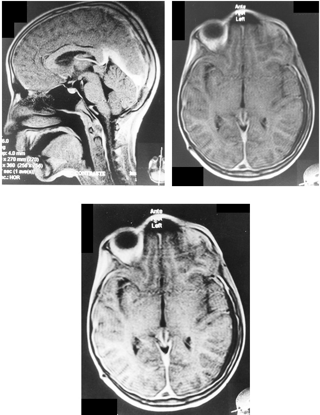
Radiological findings on axial cuts of the Computed Tomographic Scan of the head and Brain Magnetic Resonance Imaging (MRI) showed multiple lesions in the pineal, suprasellar and at the right pontine cerebellar angle (PCA) with hydrocephalus (Fig1).
A ventriculo peritoneal shunt was performed. No tissue biopsy was obtained. The analysis of Cerebro-Spinal Fluid (CSF) obtained during the ventriculo – peritoneal shunting showed the presence of tumor markers: alpha fetoprotein (αFP); and Human Chorionic Gonadotrophin (HCG). The patient then underwent radiotherapy and chemotherapy. The outcome was excellent with resolution of all the presenting symptoms and signs. The MRI done at the end of the cure revealed the remission of all the intracranial lesions (Fig2).
DISCUSSION
The development of the third ventricle leads to the displacement of the germ cell tumors outside the midline. The clinical manifestation of multiple germinomas depends on the location and size of the various tumors. Several clinical signs and symptoms are related to what is called cerebral hemiatrophy which is a result of lesions in the basal ganglia, thalamus, brainstem and cerebral hemisphere (1,4,5,7).
The radiological findings in case of intracranial germinomas are as follows: Plain X-Ray of the skull may show a calcified mass in the pineal region.
Head CT scan: the tumor usually appears as a circumscribed round or lobulated lesion, with iso- or hyperdense enhancement after contrast injection.
On brain MRI the lesion is iso-intense on T1, iso or hyper intense on T2 with homogenous enhancement after gadolinium contrast injection (1,3,9) Hydrocephalus may be present because of the stenosis of the aqueduct of Sylvius near the pineal region.
Tumors in the common location are often smaller than 60ml whereas the germinomas in atypical locations tend to be larger because of their high proliferation potential (8,9)
In atypical locations, the content of cystic part is xanthocromic; but in usual location it may be hemorrhagic. A cystic component is present in only 4% of germinomas in a normal location, 50-100% in the suprasellar location, and 83-90% in tumors located in the basal ganglia.
Analysis of CSF to determine the presence of tumor markers (ά-FP, βHCG) is important. If tumor markers are absent, then tumor biopsy must be considered. MRI of the spine also indicated for exclusion of spinal seeding which is often frequent (7,8,9) Since intracranial germinomas are malignant tumors that are sensitive to chemotherapy and radiation.
If the tumor markers are positive then radiation and /or chemotherapy is indicated (1, 6,10) In our case ,we used both of them and our results were very good.
The indication for surgical intervention especially stereotactic biopsy is still being debated, because of its high rate of mortality and morbidity (10,11) In some cases with negative tumor markers most authors recommend stereotactic biopsy for an efficient adjuvant therapeutic intervention (9,11).
Also in some cases ventriculo-peritoneal shunting is necessary to release ICP as it was performed in our case even though the risk of seeding of the peritoneum with tumor has been described in rare cases in the literature (8).
CONCLUSION
Germinomas are sensitive to radiotherapy and chemotherapy. The role of surgical treatment is still a matter of debate.


FIGURE 2: MRI T1
REFERENCES
- BIRNBAUM T, PELLKOFER H, BUETTNER U. Intracranial germinoma clinically mimicking chronic progressive multiple sclerosis. J Neurol. 2008;255(5):775-6.
- KOIZUMI H, OKA H, UTSUKI S, SATO S, TANIZAKI Y, SHIMIZU S, SUZUKI S, et al. Primary germinoma arising from the midbrain. Acta Neurochir (Wien). 2006; 148(11):1197-200; discussion 1200.
- KONNO S, OKA H, UTSUKI S, KONDOU K, TANAKA S, FUJII K. Cystic germinoma arising from the right temporal lobe. Acta Neurochir (Wien). 2002;144(8):847-8; discussion 848.
- KURIMOTO M, NISHIJIMA M, HAYASHI N, ENDO S, TAKAKU A. Multiple germinomas due to early intracranial metastasis. J Clin Neurosci. 1995; 2(1):73-5.
- LAFAY-COUSIN L, MILLAR BA, MABBOTT D, SPIEGLER B, DRAKE J, BARTELS U, HUANG A, et al. Limited-field radiation for bifocal germinoma. Int J Radiat Oncol Biol Phys. 2006;65(2):486-92.
- LAKHDAR F, HEMAMA M, LAGHMARI M, GANA R, MAAQILI R, BELLAKHDAR F. Double localization of a cerebral germinoma. Case report. J Neuroradiol. 2008; 35(3):177-80.
- MATSUMOTO K, TABUCHI A, TAMESA N, NAKASHIMA H, OHMOTO T. Primary intracranial germinoma involving the midbrain. Clin Neurol Neurosurg. 1998;100(4):292-5.
- MURRAY MJ, METAYER LE, MALLUCCI CL, HALE JP, NICHOLSON JC, KIROLLOS RW, BURKE GA.Intra-abdominal metastasis of an intracranial germinoma via ventriculo-peritoneal shunt in a 13-year-old female. Br J Neurosurg. 2011;25(6):747-9.
- SARTORI S., LAVERDA A.M., CALDERONE M., CAROLLO C., VISCARDI E. and al. Germinoma with synchronous involvement of midline and off-midline structures associated with progressive hemiparesis and hemiatrophy in a young adult. Childs Nerv Syst 2007; 23:1341-5.
- UTSUKI S, OKA H, TANIZAKI Y, KONDO K and al. Radiological features of germinoma arising from atypical locations. J Neurol 2008;255:775-6.
- Van BATTUM P, HUIJBERTS MS, HEIJCKMANN AC, WILMINK JT, NIEUWENHUIJZEN KRUSEMAN AC. Intracranial multiple midline germinomas: is histological verification crucial for therapy? Neth J Med. 2007;65(10):386-9.